These are different but complementary exams. We share the study carried out by GVM – Care & Research.
The exam electromyography is an instrumental investigation composed of two different and complementary techniques: theelectromyography, which allows you to study muscle activity, and theelectroneurographyfor the study of the peripheral nervous system. Unless otherwise indicated by the specialist, in most cases both techniques are used during the examination, in order to reach a clear diagnosis with a complete picture of the peripheral nervous system, neuromuscular activity and muscles. Below is the in-depth study for GVM – Care & Research by Dr Raffaele D’UrsiHead of Neurology at the Santa Maria di Bari Hospital.
Electromyography: for which pathologies it is indicated
As far as the study of muscles is concerned, electromyography is used in the diagnosis of both inflammatory (such as myositis) and degenerative (such as muscular dystrophies) pathologies, with symptoms that refer to muscle weakness, muscle pain, cramps. For this investigation, the electromyograph acquires the data to be analyzed by inserting a sterile and disposable needle electrode into the muscle to be examined, in order to record its activity at rest and after muscle contraction.
Electroneurography: diagnosed pathologies
The study of the nerves by means of electroneurography is used for the diagnosis of inflammatory pathologies, such as polyneuritis, both idiopathic and linked to dysmetabolic diseases such as diabetes, renal insufficiency or subsequent to the use of chemotherapy. The electromyographic examination with electroneurography is then used in the diagnosis of compression neuropathies such as carpal tunnel syndrome, cubital canal syndrome, tarsal tunnel syndrome. Peripheral nerve disorders often manifest with symptoms such as tingling, numbness, or changes in sensation. The compression of the roots of the spinal nerve – generally due to a herniated disc or a degenerative pathology of the vertebrae – configures, on the other hand, a picture of radiculopathy characterized by motor problems in one limb, associated with painful symptoms of considerable extent.
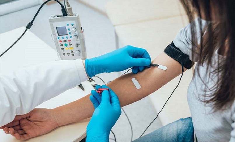
The test is performed by applying two electrodes, one for stimulation and one for recording, to the skin. The detection of the speed and amplitude of the stimulus makes it possible to evaluate the functionality of the previously identified nerve under examination. A further field of application of the electromyographic examination is the diagnosis of latent tetany syndrome, also called spasmophilia. This pathology is caused by a lack of calcium in the muscle cell and is characterized by very heterogeneous symptoms: fatigue, muscle cramps, insomnia, chest tightness, abdominal pain, anxiety.
How is the electromyography examination performed?
The electromyographic examination is preferably followed by a neurologist expert in neurophysiology, as a completion of the patient’s clinical examination during a specialist visit. The patient, based on the district to be investigated, is asked to sit or lie down on the bed. The survey takes approximately 30 minutes. At the end of the examination, which is performed on an outpatient basis, the patient is perfectly capable of immediately resuming his normal daily activity.
Preparation and contraindications
The electromyographic examination does not require any type of preparation, except the foresight to avoid the use of body creams in the imminence of the examination. It does not require preventive fasting or the suspension of any drug. The investigation is not particularly painful, although the application of the needle electrode can be annoying. The perception of discomfort is substantially subjective, and in any case resolves itself in the time it takes to carry out the examination.
There are no particular contraindications to the execution of the examination, except the absolute one of the use of the needle electrode in patients undergoing anticoagulant treatment and the one relating to the use of electrodes with current passage in patients with pacemakers or defibrillators. In the latter cases, the evaluation of the nerves pertaining to the supra-diaphragmatic districts is generally avoided.
Nurse Times editorial team
Fonte: GVM – Care & Research
Stay up to date with Nurse Times, follow us on:
Telegram – https://t.me/NurseTimes_Channel
Instagram – https://www.instagram.com/nursetimes.it/
Facebook – https://www.facebook.com/NurseTimes. NT
Twitter –
