TRENTO. New hope in the fight against brain and head and neck tumors comes from artificial intelligence, which allows for more precise and faster diagnoses.
This important frontier of innovation in medicine is now at the center of a new collaboration between the University of Trento and the Provincial Agency for health services, as a note from the Province communicates.
“With artificial intelligence and advanced medical image analysis – we read – patients’ diagnosis, treatment planning and life expectancy improve.
In collaboration with the Neuroradiology Operational Unit, interdisciplinary skills come into play: from statistics, to biomedical imaging analysis, up to computer skills in the field of augmented reality and virtual reality.
For the University of Trentino it is one of the first projects of the Inter-university Center that has started its activities in recent months and was born thanks to an intense collaboration with the University of Verona “.
This new line of collaboration. the press release continues, it involves on the one hand the University of Trento with a multidisciplinary group of researchers from various departments, and on the other the provincial health services company with personnel belonging to the Neuroradiology operating unit.
Employing artificial intelligence in place of normal diagnostic procedures has an important advantage.
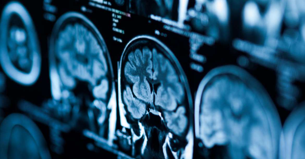
The analysis techniques, in particular, allow the quantitative and qualitative information of the tumor to be analyzed quickly and without invasive procedures, which is not always detectable through traditional visual observation.
Medical images, acquired by magnetic resonance, CT or PET, are converted by algorithms into numerical data.
This is a large amount of data to analyze: an ideal area in which the statistical techniques of artificial intelligence find application.
With the computer, numerous specific morphological characteristics of the tumor (shape, volume, tissue structure) are extracted which can be associated with the molecular and genomic characteristics of the same lesion.
This analysis will allow to evaluate the clinical risk in detail, to predict with greater precision the aggressiveness of the tumor and, consequently, to select the most suitable therapies.
The experimentation of diagnosis using artificial intelligence will start on some brain tumors (gliomas and meningiomas) and in the head and neck area.
In these types of cancer, early diagnosis is essential: if treated in advance and if the size is small, there are indeed good hopes for recovery.
If, on the other hand, the tumor is large, the prognosis will be worse and a prolonged and certainly more invasive treatment will be necessary.
Artificial intelligence thus supports the diagnostic work by neuroradiologists and that of planning the intervention for the benefit of oncologists and neurosurgeons.
The same technology will then allow to follow the clinical evolution in the patient through non-invasive devices for monitoring vital signs.
The experimentation was started with the Apss Neuroradiology Unit, engaged in recent months in an early characterization project of molecular patterns of gliomas through the analysis of magnetic resonance images and with the support of radiomics techniques.
The project is coordinated by Paola Feraco, APSS neuroradiologist and sees the involvement of the University of Bologna.
The data analysis is entrusted to a research team of the University of Trento, coordinated by Massimo Donelli, professor of electromagnetic fields at the Department of Information Engineering and Science and Giuseppe Espa, full professor of Economic Statistics at the Department of Economics and Management. .
Latest generation software will be used, which allow complete management and control of the entire process even by users with non-advanced statistical skills.
«The analysis of this type of data may also be useful for the training of new medical students and students as well as for future postgraduates and postgraduates in Radiology – explains Dr. Feraco. It will allow them to learn, with a more modern approach, a global and multidisciplinary vision of oncological pathologies, but also of other pathologies. All this with a view to increasingly developing an approach that takes into account the specificity of the patient with a treatment tailored to his needs ».
The idea of applying new technologies to this specific problem is one of the first initiatives of the newly established Center for the Sciences of Security and Crime (CSSC) of the University of Trento, whose activity is starting up in recent months.
«Collaboration with the provincial health services agency is possible thanks to teamwork between different skills. Alongside the techniques and state-of-the-art instrumentation in the statistical field, we find those of biomedical imaging analysis, skills in augmented reality, virtual reality and information technologies »comments Giuseppe Espa.
«In recent times we have witnessed a strong push towards innovation in support of medicine.
Health risks and medical risks are part of the research activities in which the University of Trento is investing a lot, because understanding and managing them requires strong multidisciplinary components, well present in our University and easily integrated thanks to the collaboration with the University of Verona. “.
«The project has only started a few months ago but we are already able to analyze a very large database for the benefit of early diagnosis – adds Massimo Donelli. We apply the data collected thanks to this software to make predictions and better characterize a particular type of tumor. We are able to obtain and make available to doctors accurate information also on the evolution of the disease and to suggest them the most suitable technique to attack it.
The idea of applying machine learning in a personalized way in this area was born through the comparison with clinicians, applying our respective skills to new problems born in the medical-health sector. In fact, at the moment there are no protocols for this type of diagnosis: our goal is to develop one thanks to the collaboration with Apss ».
The synergy in the diagnosis of these tumors is only one of the most recent activities that have seen the University and Apss work side by side. Among the most recent there is also the collaboration for the development of support for the diagnosis of type two diabetes, through a sensor of galvanic resistance at the skin level.